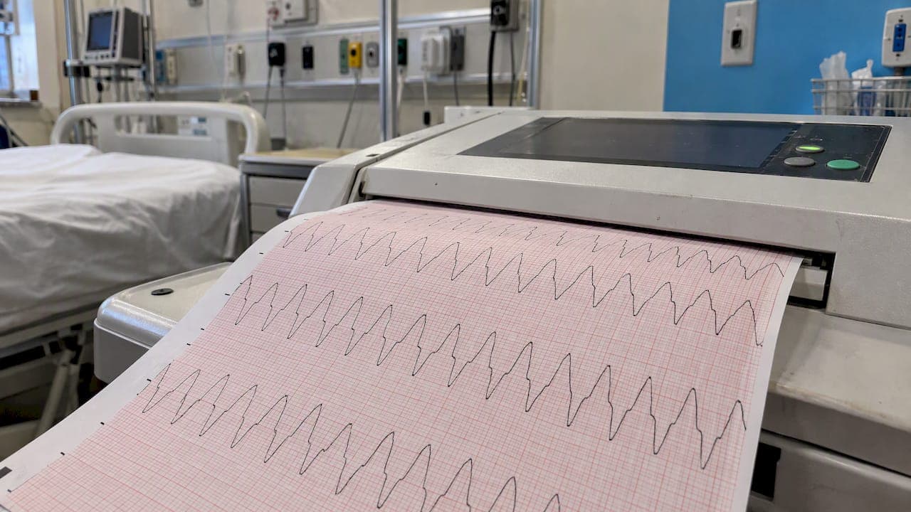
Basic EKG on the NCLEX comes down to three skills. Read a strip in the same order every time, name the handful of rhythms that get tested, and know what the nurse does first. You don't need to be a cardiologist. You need a routine that works under time pressure and the judgment to pick the safest action.
This guide gives you the routine, the rhythm groups, the numbers worth memorizing, the priority rules for dangerous rhythms and a practice plan you can start today.
TL;DR
- Read every strip in the same order: rate, rhythm, P waves, PR interval, QRS width, then the patient. Never treat the monitor before you look at the person.
- Paper speed is 25 mm/s, so one small square is 40 ms and one large square is 200 ms. Those two numbers drive every rate and interval measurement.
- Ventricular fibrillation and pulseless ventricular tachycardia are shockable. Asystole and pulseless electrical activity are not.
- Questions usually ask what you do first. Assess the patient, check for a pulse, then act on the rhythm.
- Short daily practice with rationales beats long weekend cramming for pattern recognition.
Table of Contents
- What the NCLEX actually expects from you
- A six-step routine for reading any strip
- The rhythms you are most likely to see
- Shockable versus nonshockable: the rule that earns points
- Intervals worth memorizing
- When the strip lies: artifact and technique
- How to choose the safest answer
- A worked example
- A study plan that sticks
- Practice EKG questions every day with RecallOS
- FAQ
- Sources
What the NCLEX actually expects from you
The NCLEX is not an arrhythmia certification exam. The 2026 NCLEX-RN Test Plan, effective April 2026, places performing electrocardiograms within the nurse's scope, along with assessing changes in vital signs and monitoring diagnostic test results and changes in client condition. In practice, that means the exam wants to know whether you can:
- Recognize a rhythm that is normal, a rhythm that needs monitoring and a rhythm that needs action now.
- Connect the strip to the patient: pulse, blood pressure, level of consciousness, chest pain, shortness of breath.
- Choose the next nursing action in the right order.
- Prepare the patient and equipment correctly when an EKG is ordered.
If you want the bigger picture of how the exam is built, our breakdown of the NCLEX test plan shows where cardiac content sits among the client need categories. Rhythm questions are also a natural fit for Next Gen item types, where you read a case, notice the cues and decide what to do. Our guide to Next Gen NCLEX strategies explains how to practice that reasoning.
A six-step routine for reading any strip
The trick is to stop looking at the whole squiggle and start asking the same questions in the same order. A routine removes panic, and panic is what costs points on this topic.
Step 1: Know your grid
Standard EKG paper runs at 25 mm/s. According to a technologist's guide to electrocardiography, one small square represents 40 ms and one large square represents 200 ms. Five small squares make one large square, so five large squares equal one second.
That gives you two quick rate methods:
- Regular rhythm: count the large squares between two R waves and divide 300 by that number. Three large squares is about 100 beats per minute. Five is about 60.
- Irregular rhythm: count the R waves in a six-second strip and multiply by 10.
The 300 divided by large squares shortcut follows directly from the 200 ms large square, because 60 seconds divided by 0.2 seconds is 300.
Step 2: Is the rhythm regular?
Look at the distance between R waves. Are they evenly spaced, slightly varying or completely chaotic? Regular, regularly irregular and irregularly irregular are three different clues. Chaotic spacing with no clear P waves points you toward atrial fibrillation (if the QRS complexes are narrow and the patient is perfusing) or toward something much worse if the baseline itself is chaotic.
Step 3: Are there P waves, and does each one have a QRS?
Look for one upright P wave before every QRS. A P wave in front of every QRS with a consistent shape suggests the impulse starts in the sinus node. Missing P waves, extra P waves or P waves that march along independently of the QRS tell you the problem is in the atria or the conduction between atria and ventricles.
Step 4: Measure the PR interval
The PR interval runs from the start of the P wave to the start of the QRS. It reflects the time the impulse takes to cross the AV node. Use your course reference for the normal range and memorize it. For the exam, you need to recognize three patterns: a consistently long PR, a PR that lengthens until a beat drops, and a PR with no relationship to the P waves at all.
Step 5: Look at the QRS width
A narrow QRS means the impulse traveled down the normal conduction pathway. A wide, bizarre QRS means the ventricles are depolarizing in a slower, abnormal way, which is why ventricular rhythms look wide. Width is often the fastest way to split rhythms into "probably supraventricular" and "possibly dangerous."
Step 6: Check the patient
This is the step most students skip, and it is the step the exam cares about most. Before you decide anything, ask: is there a pulse, is the patient awake, what is the blood pressure, is there chest pain, dyspnea or dizziness? A lethal-looking strip on a patient who is chatting and brushing their teeth is more likely a loose lead than a cardiac arrest.

The rhythms you are most likely to see
Rather than memorizing dozens of strips, sort them into families. Each family has one defining feature and one typical nursing priority.
| Family | Examples | What defines it | What the question usually asks |
|---|---|---|---|
| Sinus rhythms | Normal sinus rhythm, sinus bradycardia, sinus tachycardia | P before every QRS, regular, differing only in rate | Find the cause (pain, fever, hypovolemia, medications) and assess tolerance |
| Atrial rhythms | Atrial fibrillation, atrial flutter | No distinct P waves with an irregularly irregular rhythm, or sawtooth flutter waves | Assess perfusion, anticipate rate control and stroke risk teaching |
| AV blocks | First-degree, second-degree types I and II, third-degree | Problems in PR interval or P-to-QRS relationship | Recognize which block is more dangerous and when to expect pacing |
| Ventricular rhythms | PVCs, ventricular tachycardia, ventricular fibrillation | Wide QRS, no organized P waves in the dangerous forms | Pulse check first, then the arrest pathway |
| Arrest rhythms | Asystole, pulseless electrical activity | No effective mechanical output | CPR, epinephrine and reversible causes |
Sinus rhythms: the baseline
If you can recognize normal sinus rhythm confidently, every abnormal rhythm becomes a deviation from something you know. Sinus bradycardia and sinus tachycardia share the same shape and differ only in rate. The exam rarely asks you to treat the number. It asks you to figure out why the rate changed and whether the patient tolerates it.
Atrial fibrillation and flutter
Atrial fibrillation has no organized P waves and an irregularly irregular ventricular response. Atrial flutter shows a regular sawtooth baseline. In both, the nursing questions tend to focus on perfusion, rate control, anticoagulation and symptoms of embolic events. Stay with the patient assessment first: a stable patient and an unstable patient with the same strip get different answers.
AV blocks: a pattern, not a formula
Think of the AV node as a gate between atria and ventricles.
- First degree: every P gets through, just slowly. The PR is consistently long.
- Second degree, type I: the PR gets longer and longer until one P wave fails to produce a QRS, then the cycle restarts.
- Second degree, type II: the PR stays constant, but some P waves suddenly fail to conduct.
- Third degree: atria and ventricles beat independently. P waves and QRS complexes have no relationship.
The higher the degree, the more the exam cares about symptoms such as dizziness, hypotension and syncope, and about the need for pacing. Always confirm the patient's tolerance before choosing an action.
Ventricular rhythms
Premature ventricular contractions appear as early, wide beats. Occasional PVCs are common and usually prompt assessment rather than emergency treatment, but frequent or multifocal PVCs in a patient with symptoms or new changes get reported. Ventricular tachycardia is a fast, wide-complex rhythm. Ventricular fibrillation is chaotic with no identifiable QRS.
The difference between a patient with ventricular tachycardia who has a pulse and one who does not is the difference between urgent treatment and the cardiac arrest algorithm. The pulse decides it.
Shockable versus nonshockable: the rule that earns points
American Heart Association algorithms classify ventricular fibrillation and pulseless ventricular tachycardia as shockable rhythms that require defibrillation along with CPR. Asystole and pulseless electrical activity are nonshockable. They are managed with CPR, epinephrine and treatment of reversible causes rather than defibrillation.
A simple memory frame:
- Chaos with no pulse (VF, pulseless VT): shock.
- Flat line or organized rhythm with no pulse (asystole, PEA): do not shock. Compress, give epinephrine and look for the cause.
The 2020 AHA guidelines for adult advanced cardiovascular life support stress that high-quality CPR is critical, with chest compressions at a rate of 100 to 120 per minute and a depth of at least 2 inches. The same guidance calls defibrillation critical for cardiac arrest from ventricular fibrillation or pulseless ventricular tachycardia.
After return of spontaneous circulation, the AHA post-cardiac arrest pathway says early evaluation of a 12-lead ECG and hemodynamics informs decisions about cardiac intervention, including when STEMI is present. Expect a question that asks what to obtain after a patient regains a pulse.
Emergency skills outside the rhythm strip often share a basic life support mindset. If you want a refresher on a different emergency sequence, this walkthrough of infant choking response steps is a quick one to read alongside your CPR review.
Intervals worth memorizing
You do not need an interval table full of numbers, but you do need the few that are asked directly and the clinical meaning behind them.
- Grid values: 40 ms per small square, 200 ms per large square at 25 mm/s.
- QTc: a normal corrected QT interval is less than 440 ms. A prolonged QT matters because it raises the risk of dangerous ventricular rhythms, so questions often pair it with medication review, electrolyte levels and the instruction to report it.
- PR interval: reflects AV node conduction time. A long PR points toward AV block patterns.
- QRS width: wide complexes suggest a ventricular origin or abnormal conduction.
Electrolytes show up in cardiac questions constantly, because potassium and other levels change the strip. Pair this topic with our fluids and electrolytes cheat sheet so the two reinforce each other.
When the strip lies: artifact and technique
Not every ugly tracing is a dangerous rhythm. Research on EKG technique notes that poor skin preparation or inadequate contact can weaken the cardiac signal and raise skin impedance, and that muscle tremor or movement can create artifacts.
For the exam, this translates into a clear priority order:
- Look at the patient. Are they awake, breathing and perfusing?
- Check the pulse.
- If the patient looks fine and the strip looks terrible, check the leads, skin contact and movement before calling anything an emergency.
- If the patient looks unwell, do not waste time on the leads. Treat the patient and call for help according to protocol.
The same source also makes a point worth remembering: ECG interpretation is on an individual patient basis, not by comparison between patients. On the exam, that means the patient's own baseline and symptoms outweigh any generic rule.
How to choose the safest answer
Most basic EKG questions are won by reasoning, not by recall. Try this filter on every item.
- Assess before you act. If an option says to check the patient, a pulse or perfusion and another says to give a medication or start a procedure, the assessment usually comes first unless the scenario already shows the patient is in arrest.
- Pulse or no pulse? This single fact splits the algorithm. Pulseless means CPR and the arrest pathway. A pulse means you have time to assess and treat the cause.
- Stable or unstable? Chest pain, hypotension, altered mental status and shortness of breath change the answer.
- Shockable or not? Only VF and pulseless VT get a shock.
- Escalate when the patient changes. New symptoms, a new rhythm or a worrying QTc go to the provider following your facility's protocol.
- Avoid options that delay care. Repeating a test, documenting first or waiting and watching rarely beat a direct, safe nursing action when the patient is deteriorating.
For a deeper look at this style of thinking, read our guide to NGN-ready test-taking strategies.
A worked example
A client on a telemetry unit has a monitor alarm. The strip shows a wide, fast, regular rhythm. Which action comes first?
Walk it through:
- The rhythm looks ventricular, but the strip alone is not enough.
- You go to the bedside. The client is sitting up, alert, and says the monitor beeped while they were brushing their teeth.
- You check the leads and see one has come loose, and the rhythm in the other lead looks normal.
The lesson: the answer to "what do you do first" was to assess the client, not to defibrillate or call a code. If the same client had been unresponsive and pulseless, the answer would flip immediately to starting CPR and following the shockable rhythm pathway. Same strip, different patient, different answer.
A study plan that sticks
Rhythm recognition is pattern recognition, and patterns fade without repetition. A plan that works for most students:
- Daily, 10 to 15 minutes: a handful of rhythm questions with rationales. Short, frequent sessions build recall better than one long weekly block.
- Weekly: group your misses. If most errors come from AV blocks, spend the next few days there only.
- Always: say the six steps out loud as you read a strip until they become automatic.
- Link to the case: for every rhythm, write one sentence on what the patient looks like and one on the first nursing action.
- Mix topics: rhythm questions can appear next to electrolytes, medications and emergencies, so practice them interleaved. Our NCLEX practice question guide shows how to structure that practice, and cardiac rhythm basics goes deeper on the foundation.
Our perspective
We build RecallOS with practicing nurses and CRNAs, and the most common thing they tell us about EKG studying is that students try to memorize strips instead of learning a routine. A strip you have seen once is easy to forget. A six-step habit you have repeated for weeks is hard to lose.
That is why we designed daily high-yield practice around short sessions with instant rationales. We believe the rationale matters as much as the question, because it teaches why the safest action is the safest action.
We also think weak spots deserve more of your time than strengths. Our approach is to bring the rhythms you miss back for review and let you upload your own notes so the practice reflects the material your program actually teaches. Retention is the real exam skill, and we built the tool around it.
Caleb
Practice EKG questions every day with RecallOS
If you want a steady routine instead of a pile of PDFs, the Daily Recall Run gives you a focused set of exam-style questions each day, with rationales and a streak to keep you consistent. Our design aims to bring back the topics you miss, so rhythm recognition gets repeated before it fades.

You can also upload your own notes and turn your cardiac lecture slides into flashcards and quizzes. Start with the NCLEX study app page to see how daily practice fits your exam date, or see our adaptive nursing quiz app for how weak-area review works. Pricing and plan details are in the app and on our subscription terms.
FAQ
How much EKG do I need to know for the NCLEX?
You need solid basics: reading a strip with a consistent routine, recognizing the common rhythm families and knowing the first nursing action for each. The exam focuses on safe decisions, so patient assessment matters as much as rhythm names. Review the 2026 test plan to see where cardiac content fits.
What is the fastest way to tell shockable rhythms from nonshockable ones?
Check the pulse first. In a pulseless patient, ventricular fibrillation and pulseless ventricular tachycardia are shockable, while asystole and pulseless electrical activity are not. Nonshockable rhythms are managed with CPR, epinephrine and treatment of reversible causes.
Do I need to calculate heart rate by hand?
Yes, be ready for it. At 25 mm/s a large square is 200 ms, so for a regular rhythm you divide 300 by the number of large squares between R waves. For an irregular rhythm, count R waves in six seconds and multiply by 10.
What should I do if the strip looks dangerous but the patient looks fine?
Assess the patient and check the pulse before anything else. Loose leads, poor skin contact and patient movement can all produce artifacts that mimic serious rhythms. Fix the contact, recheck the tracing and follow your facility's protocol if the patient is symptomatic.
Can I practice EKG questions daily without feeling overwhelmed?
Yes. Ten to fifteen minutes of focused questions with rationales is enough to build recognition over weeks. Keep a list of the rhythms you miss and revisit them first.
Sources
- NCLEX-RN Test Plan (NCSBN, effective April 2026)
- Electrocardiography: A Technologist's Guide (Journal of Nuclear Medicine Technology)
- 2020 AHA Guidelines for CPR and ECC: Adult ACLS
- AHA Adult Cardiac Arrest Algorithm
- AHA Post-Cardiac Arrest Care Algorithm
- Infant choking response steps